
May 28, 2021
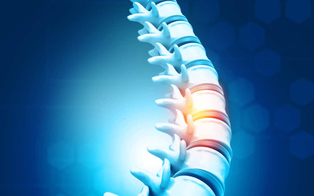
More than 80 percent of adults will experience lower back pain at some point in their lives.1 It is the single most common cause of disability worldwide 2 and the second most common cause of adult disability in the United States. 3
Most cases of lower back pain have a benign clinical course which resolves with conservative care such as rest, activity modification, anti-inflammatories, heat or ice therapy, and physical therapy. Some, however, will develop chronic low back pain or pain that lasts for 3 months or longer.
Common Causes of Chronic Lower Back Pain
- Arthritis. Osteoarthritis of the spine is a condition where there is degeneration of the joints, discs, and bones in the spine as people age. Arthritis can lead to bone spurs which result in the narrowing of the space around the spinal nerves and spinal cord, a condition called spinal stenosis.
- Facet joint dysfunction: This occurs when the facet joints, the small joints that connect the individual bones in the spinal column, get inflamed or when the cartilage in the joints becomes injured or worn out. They can also get out of normal alignment, a condition called spondylolisthesis.
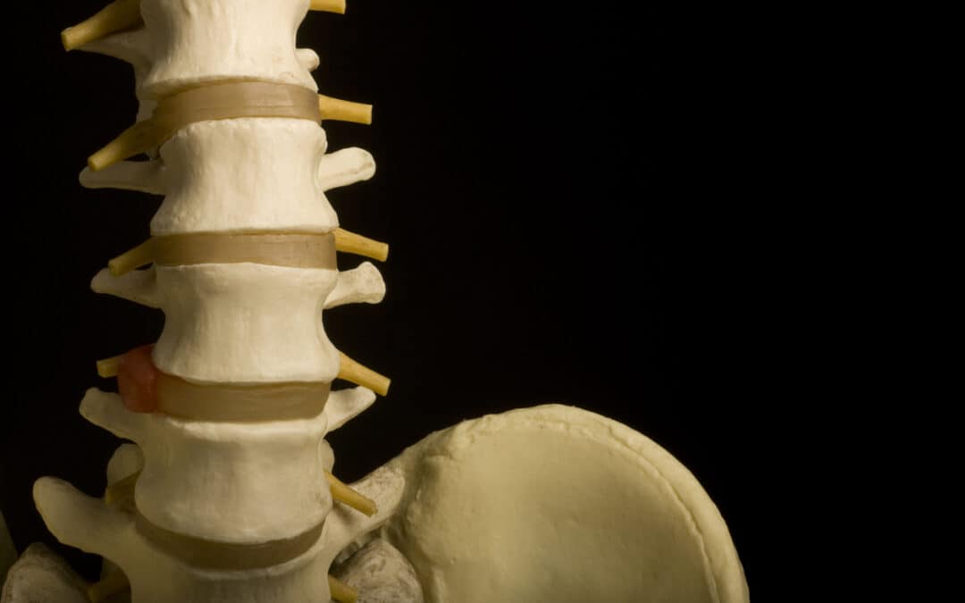
- Bulging or Ruptured Disc: The discs are soft, gel-filled structures that act as a cushion or shock absorbers between the vertebral bones in your spine. When these discs become injured, they can bulge out and can put pressure on neighboring nerves causing pain. As people age, discs lose their hydration and wear down leading to degeneration. Degenerative discs can develop tears and collapse which causes low back pain, muscle spasm, or spinal stenosis.
Other less common causes of low back pain include spinal cord problems, scoliosis, fracture, and non-spine sources like tumors and kidney stones. It is important that your physician perform a comprehensive evaluation of your medical history and physical examination to give you an appropriate diagnosis for your back pain.
Alternative Treatments
Patients who have failed standard treatments or are seeking alternatives to spine surgery for back pain should consider Regenerative therapies. These advanced treatments involve the use of your own cells to help your body heal faster. They avoid the risks and long-term complications associated with surgery and reduce pain to help you maintain an active lifestyle. Read about these regenerative treatment solutions here.
Sources:
- Sauver, JL et al. Why patients visit their doctors: Assessing the most prevalent conditions in a defined American population. Mayo Clinic Proceedings, Volume 88, Issue 1, 56–67.
- Hoy D, March L, Brooks P, et al The global burden of low back pain: estimates from the Global Burden of Disease 2010 study Annals of the Rheumatic Diseases Published Online First: 24 March 2014. doi: 10.1136/annrheumdis-2013-204428
- From the Centers for Disease Control and Prevention, Prevalence of disabilities and associated health conditions among adults—United States, 1999. JAMA 2001;285 (12) 1571- 1572

May 21, 2021
Osteoarthritis is the most common type of arthritis affecting 32.5 million Americans. It is a chronic disease that slowly degenerates the protective cartilage in your joints. For some people, symptoms are minimal. While for others, it can be disabling. The joints in the knees, hips, hands and spine are typically involved, although any joint in the body can be affected. Signs and symptoms of osteoarthritis vary, but there are early signs to watch out for that can help prompt for an early medical evaluation and subsequent treatment.
Early Warning Signs of Osteoarthritis
- Joint Pain: Pain typically worsens with activity or at the end of the day. Pain can be felt as a generalized ache in the joint or sharp sensation with certain motions.
- Swelling: Fluid accumulation inside the joint or inflammation of soft tissue around the joint can cause swelling.
- Joint Stiffness: Stiffness is typically felt early in the morning upon waking up or after a period of inactivity. The stiffness gets better after moving around and as the day progresses.
- Loss of Flexibility: You may lose full range of motion and can find it difficult to move the joint because of pain, swelling or stiffness.
- Abnormal Sensations: Popping, clicking or cracking sounds may be heard and grating sensations may be felt in the joint.
If you experience these symptoms regularly, seek consultation with your orthopedic doctor. A comprehensive medical evaluation and appropriate diagnostic testing by a board-certified physician is needed to accurately diagnose the cause of your joint pain. Read our blog and watch our webinar on the non-surgical treatment options and innovative therapies for osteoarthritis.

May 15, 2021
The intervertebral discs are gel filled structures in the spine that act as cushion in between the vertebral bones. Degenerative disc disease (DDD) is a common condition that results from wear and tear of the intervertebral discs. As we age, the discs deteriorate due to repetitive stress, increased load, and decrease in blood flow and nutrition to the discs. Degenerative changes in the discs include: annular tear (tear in the outer fibrous ring of the disc), herniation or bulge, decrease in height, drying out, stiffness and bone spurs.
Symptoms of Degenerative Disc Disease
The symptoms of degenerative disc disease are dependent on the disc that is involved and changes in the surrounding spinal structures as a result of, or associated with the degeneration. The most common symptom of degenerative disc disease is a continuous deep pain in the neck, midback or low back that occasionally flares up to a more intense, disabling pain. The episodes can last for a few days or several weeks, and typically recurs. The baseline pain is variable in individuals and can range from almost no pain, nagging ache or severe pain.
The pain typically gets worse with prolonged sitting, standing and walking. Twisting and bending the spine, or carrying heavy loads can also aggravate the pain in the back. Prolonged tilting of the head down while reading or working on the computer can worsen neck pain.
The pain can refer to the buttocks and hips when the lumbar spine is involved, or to the shoulder blade region when the cervical spine is involved. When the degenerated disc compresses or irritates the nearby spinal nerves, radiating pain can be felt in the buttocks, hips, legs and feet (lumbar disc degeneration); or in the shoudler, arm and hand (cervical disc degeneration). Individuals with disc problems can also experience muscle tightness or muscle spasms.
It is not uncommon to see degenerative changes in the spinal imaging of patients over 60 years old. Majority of these patients will not present with symptoms.These imaging findings must be interpreted in the context of the patient’s clinical condition.
Treatment Options for Degenerative Disc Disease
Nonsurgical Treatments Options
Majority of patients who develop symptoms from degenerative disc disease respond well to conservative treatments without the need for surgical intervention.
- Medications: Non-steroidal anti-inflammatory medications (NSAIDS) decrease the symptoms of pain and inflammation associated with this condition. Analgesic medications or muscle relaxants can be helpful when the symptoms are severe. Medications to decrease nerve-related pain, called “sciatica” in the lumbar spine, can also be beneficial.
- Physical Therapy: Therapeutic exercises to strengthen the supporting muscles, improve spinal alignment, flexibility and stability, are critical in healing the spine. A physical therapist can help teach proper exercises and educate on activity modifications to help prevent recurrence of symptoms and injuries.
- Regenerative Treatments: These are innovative, non-surgical treatments using cells from your own body to heal and repair orthopedic conditions. These cell therapies, called Platelet-rich plasma and Cell-based Therapies derived from Bone Marrow, have been shown to help treat spinal conditions and provide long-term relief.
Surgical Treatment Options
Surgery is an option when all conservative treatment options have failed, when there is severe, debilitating pain that interferes with daily function, or when there is worsening neurologic function. These surgeries may involve the removal of a bulging portion of the disc (discectomy), removal of a portion of the bone in the spinal canal (laminectomy), replacing the degenerated disc with an artificial disc, or spinal fusion.
For help with your back pain and degenerative disc disease, please contact the experts at San Diego Orthobiologics Medical Center.
Apr 16, 2021
Facet joints are small joints that connect the bones of the spine called vertebrae. They come in pairs on each side of the vertebral bones. They allow for motion of the spine like bending and twisting. The orientation of the facet joint dictates the direction of the motion it allows.
There are various causes of pain coming from the facet joint. Similar to the joints in your arms and legs, the facet joints are prone to wear and tear. They can develop degenerative changes or joint osteoarthritis. This is a common condition in older people. The capsule surrounding the facet joint, which is made of ligaments and connective tissue, can be overstretched creating a sprain injury. Autoimmune or Inflammatory conditions like rheumatoid arthritis can also affect the facet joints by causing inflammation and deterioration.
Pain in the facet joints are typically felt in the local region of the affected joint segmetns. The pain can be felt close to the midline in the neck, upper back, mid-back or low back. Sometimes the pain is referred to the head, shoulder blade, shoulder, thigh or buttock. Stiffness, grinding in the joints (crepitus) and muscle spasm are other symptoms associated with facet joint disorders.
The standard treatment for facet joint pain include a course of physical therapy, non-steroidal anti-inflammatory medications, pain relieving medications, and home exercise program. When conservative measures fail, injection therapy to the facet joints or to the nerves leading to the facet joints can be helpful.
What is a Facet Joint Injection?
Facet joint injection is a minimally invasive procedure that is performed in an outpatient medical clinic. Local anesthesia is typically used in the procedure. There is no need for sedation in most cases. The procedure is performed under fluoroscopic (live digital x-ray) guidance. The typical substances that are injected to the facet joint are steroid medication to decrease inflammation and local anesthesia to provide immediate pain relief. The effects of these medications can provide temporary pain relief that typically last for 3-4 months.
Another injection that can provide pain relief does not involve injection to the joint, rather it involves the injection of numbing agents to the pain transmitting nerves (medial branch) called a nerve block. If this procedure results to a positive reponse, another procedure called Radiofrequency Ablation (RFA) can be performed. This involves inducing a heat lesion to the medial branches to prevent transmission of pain signals to the brain.
The use of Orthobiologics is an alternative treatment to facet joint conditions. Although the research on these treatments for spinal conditions is relatively new, it has the potential to improve arthritis and provide long term pain relief.
Risks Associated with the Facet Joint Injection Procedure
Facet joint injections are relatively safe with minimal risks. Risks associated with this procedure may include:
- Discomfort at the Injection Site: These effects are temporary and typically resolve in a few hours.
- Adverse reaction to medications: Some of the medications used in the procedure can cause adverse reaction in susceptible individuals. It is important to review your medications with your physician prior to the procedure. Severe allergic reactions are rare.
- Bleeding: Patients who are taking blood thinners or have a bleeding disorder are at risk for bleeding when undergoing an injection procedure. Blood thinners are typically stopped for 3-5 days prior to the procedure, with the permission of your primary doctor, to avoid this risk.
- Infection: Infection is rare when the procedure is performed under strict sterile protocols and proper procedure technique.
Sources:
https://www.ncbi.nlm.nih.gov/pmc/articles/PMC1905869/
http://www.ajnr.org/content/33/8/1419
https://www.ncbi.nlm.nih.gov/pmc/articles/PMC6206372/
Apr 2, 2021
Epidural steroid injection (ESI) is a minimally invasive procedure to help reduce inflammation and pain caused by spinal nerve root irritation. The spinal nerve can be irritated by compression caused by herniated discs, bone spurs, or narrowing of the spinal canal otherwise known as spinal stenosis. Pain, tingling, numbness, or weakness along the nerve distribution in the arms or legs are typical symptoms and are referred to as radiculopathy. This pain can persist for years if left untreated.
The goal of an epidural steroid injection is to help reduce inflammation along the nerve root. The procedure aims to reduce pain so patients can return to normal activity and participate in a rehabilitation program to strengthen the spine.
What is an Epidural Steroid Injection?
Epidural Steroid injections (ESI) involve injecting a solution of medication in the area between the bone and the protective layer of the spinal nerve called the epidural space. The medications consist of a potent anti-inflammatory called corticosteroids and a numbing agent like lidocaine.
Epidural steroid injections may be performed in the neck (cervical), middle back (thoracic), or lower back (lumbar). The injection is performed through the nerve canals on the side of the spine (transforaminal epidural injection) or through the middle of the spine (interlaminar epidural steroid injection).
What are the indications for this Injection?
Epidural steroid injections are used in patients who suffer from pain in the neck, mid-back, or low back which can radiate to the arms or legs. Patients who have the following conditions are candidates for epidural steroid injections:
- Spinal Stenosis – narrowing of the spinal canal and nerve root canal.
- Herniated Disc – the cushion between bones in the spine, called the intervertebral disc, can bulge or rupture through small tears and cause inflammation in the nearby spinal nerve.
- Degenerative Disc – breakdown or aging of the spinal disc can cause a collapse of the disc, formation of bone spurs, and narrowing of the spinal canal.
- Radiculopathy– irritation of spinal nerves due to nerve canal narrowing, disc bulge, or compression which causes pain to travel into the arms or legs, also known as “sciatica”.
An epidural steroid injection can also be used to determine whether surgery will be successful in patients who have a herniated disc.
Risks Involved with Epidural Steroid Injections
ESI procedures have been performed for many years and are considered safe and effective. Although uncommon, some patients may experience temporary side effects from the steroid medication in the form of facial flushing, insomnia, or irritability.
Serious complications are very rare, which may include an allergic reaction to the medications or contrast dye used, infection, nerve damage, or bleeding. When this procedure is performed properly with fluoroscopic guidance, the risks are minimized. This procedure is typically well tolerated by most patients and provides immediate relief that may last for up to 6 months.
For patients who are unable to obtain long-lasting pain relief or who wish to avoid the use of steroids, natural treatment options exist. These treatments use your own cells, helping with pain, and even may heal your injury. For more information, visit our website to learn more about PRP and cell therapy treatments.
Feb 5, 2021
The researchers and scientists of the International Human Genome Project first mapped our full genetic blueprint 18 years ago.[i] Their breakthrough efforts enable numerous advances in how we treat and prevent various diseases and orthopedic conditions.
The Human Genome roadmap allows us to better understand the genes that are associated with certain diseases and how these genes can predispose a person to develop these diseases. It can also help in understanding how genes can be altered to improve disease processes.
In the field of orthopedics, a group of scientists at Stanford University discovered a biological indicator of cartilage degeneration and inflammation in patients with arthritis, disc degeneration, and sciatica. This molecular complex is called the Fibronectin-Aggrecan Complex or FAC.
Alpha-2-Macroglobulin (A2M) is a Master Protease Inhibitor
Having identified the FAC molecular complex, the Stanford team recognized how this biomarker could be used in conjunction with a naturally-occurring blood protein called Alpha-2 Macroglobulin or A2M to improve the effectiveness of treatments for degenerative disc disease and joint arthritis. The FAC can be measured to predict which patients will have the best results with A2M therapy.
A2M inhibits the breakdown of collagen in cartilage and has been scientifically proven to stop the progression of joint cartilage and disc degeneration. It is known as a Master Protease Inhibitor. It inhibits proteases, which are enzymes that breaks down proteins in cartilage. Much like a levee along a rising river, it “holds back” and prevents damage to tissues.
How A2M therapy Works
A2M therapy is a non-invasive treatment performed on an outpatient basis. After conducting a complete examination, your physician will determine if you are a good candidate for this treatment. Further imaging studies such as x-ray, ultrasound, or MRI may be necessary to determine the cause of your pain.
The A2M procedure starts with a simple blood draw from the arm. The treatment is autologous, which means the A2M is derived from the patient’s own body. There are no donor products used. The blood is concentrated using an FDA-approved device in our lab. After numbing the treatment area, the concentrated A2M is injected on the same day. Digital x-ray or ultrasound guidance will be used to make the treatment comfortable, safe and precise.
Currently, this breakthrough treatment is only being offered by a select number of physicians across the country. By screening patients for these treatments, regenerative medicine specialists are able to offer targeted A2M treatment to appropriate patients. This gives patients new hope for relief of pain and inflammation due to common orthopedic conditions.
Drs. Christopher Rogers and Mary A. Ambach of San Diego Orthobiologics Medical Group are two of just a handful of physicians in San Diego who offer A2M therapy for their patients. They have found this and other new regenerative medicine therapies valuable for the treatment of pain and inflammation associated with arthritic joints and disc degeneration.
Sources:
[i] https://www.genome.gov/human-genome-project#:~:text=Beginning%20on%20October%201%2C%201990,is%20the%20Human%20Genome%20Project%3F
https://cytonics.com/
https://sdomg.com/master-protease-inhibitor-a2m/