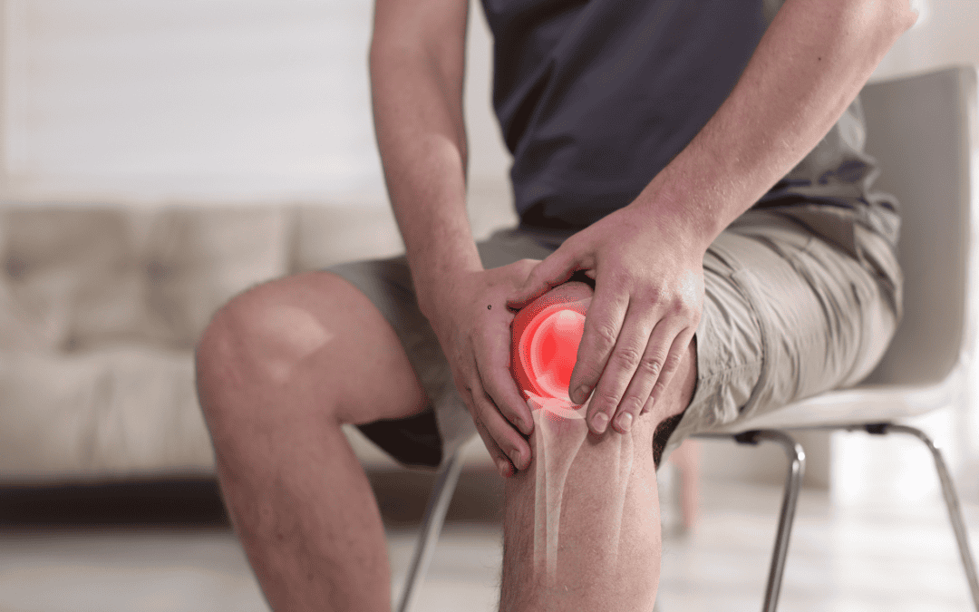
Chronic joint, tendon, and ligament pain can make everyday activities feel like a challenge. When conservative treatments like rest, physical therapy, and anti-inflammatory medications are not enough, injection therapies are often the next step. Two of the most common options are Platelet-Rich Plasma (PRP) therapy and cortisone (steroid) injections.
While both treatments are used to relieve pain and inflammation, they work in very different ways. Understanding the differences can help you make an informed decision about your care.
What Is PRP Therapy?
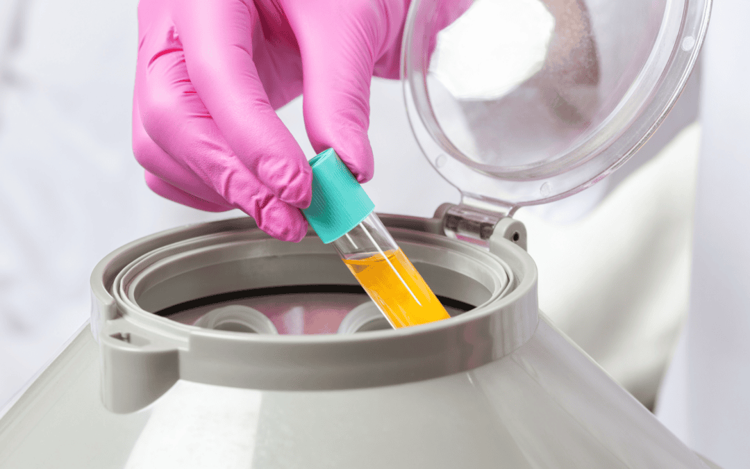
Platelet-Rich Plasma (PRP) therapy is a regenerative treatment that uses your body’s own healing properties. A small sample of your blood is drawn and placed in a centrifuge to separate and concentrate the platelets. These platelets contain growth factors that help support tissue repair.
The concentrated PRP is then injected into the injured or painful area, such as:
Knee osteoarthritis
Tendon injuries (like tennis elbow or Achilles tendonitis)
Shoulder pain
Hip pain
Mild ligament injuries
PRP focuses on promoting healing at the source of the problem rather than simply masking symptoms.
What Are Cortisone Injections?
Cortisone injections contain a powerful anti-inflammatory medication (a corticosteroid). When injected into a joint or soft tissue, cortisone quickly reduces inflammation and swelling, often providing rapid pain relief.
Cortisone injections are commonly used for:
Arthritis flare-ups
Bursitis
Tendinitis
Acute joint inflammation
Spinal joint irritation
They are especially helpful when inflammation is severe and immediate symptom relief is needed.
How They Work: Regeneration vs. Suppression
The biggest difference between PRP and cortisone lies in how they treat pain.
PRP:
Stimulates tissue healing
Encourages cellular repair
May improve tissue quality over time
Often used for chronic or degenerative conditions
Cortisone:
Reduces inflammation quickly
Suppresses the immune response
Primarily addresses symptoms
Often used for acute inflammation or flare-ups
PRP aims to repair. Cortisone aims to calm inflammation.
Speed of Relief
Cortisone injections often provide relief within a few days. This makes them appealing for patients who need fast improvement, such as before an event or during a severe flare-up.
PRP therapy typically takes longer to show results. Some patients notice improvement within a few weeks, but full benefits may take several weeks to months as the tissue heals.
Duration of Results
Cortisone relief may last weeks to several months, but repeated injections can potentially weaken tissues over time.
PRP results may take longer to appear but can last longer because the treatment supports healing rather than temporary suppression.
Safety and Side Effects
Both treatments are generally safe when performed by experienced providers.
Cortisone risks may include:
Temporary pain flare after injection
Tissue weakening with repeated use
Elevated blood sugar (important for diabetic patients)
PRP risks may include:
Temporary soreness at the injection site
Mild swelling
Minimal risk of allergic reaction since it uses your own blood
Because PRP uses your body’s natural components, it carries a low risk of adverse reactions.
Which Treatment Is Right for You?
The right choice depends on several factors:
Severity of pain and inflammation
Treatment goals (short-term relief vs. long-term healing)
Other health issues
Previous treatment history
In some cases, cortisone may be appropriate to quickly calm severe inflammation before beginning a longer-term strategy like PRP and rehabilitation.
Personalized Care at SDOMG
At SDOMG, treatment decisions are based on your specific diagnosis, activity level, and long-term goals. Whether you need fast relief from a painful flare-up or a regenerative approach to support tissue healing, we help guide you toward the option that best supports your recovery.
If you are experiencing persistent joint or tendon pain, schedule a consultation to discuss whether PRP therapy, cortisone injections, or another treatment approach is right for you.
Your recovery plan should be as individualized as you are.
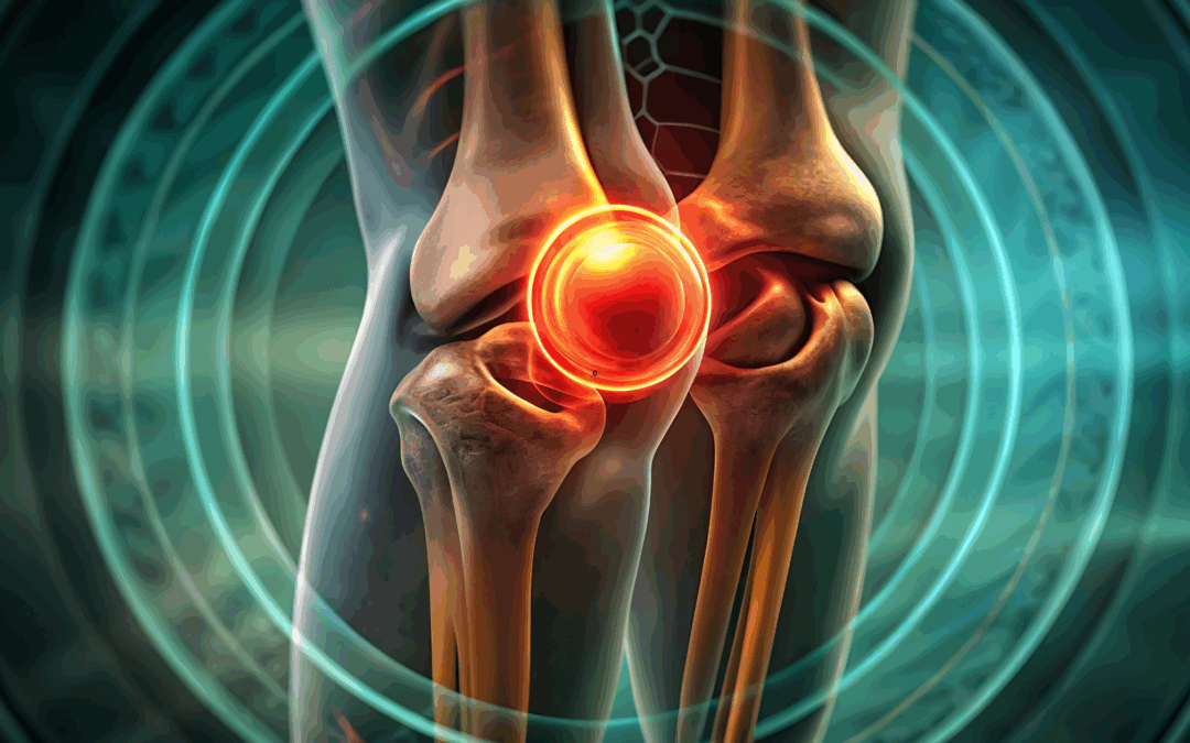
Joint preservation focuses on protecting and maintaining the natural function of joints for as long as possible. Instead of immediately resorting to joint replacement surgery, joint preservation aims to slow or halt joint degeneration, reduce pain, and improve mobility through early diagnosis and targeted treatments.
As arthritis and joint degeneration often develop gradually, early intervention plays a critical role in maintaining joint health. By addressing symptoms and structural changes early, patients may be able to delay—or even avoid—the need for surgical joint replacement.
What Causes Early Joint Degeneration?
Joint degeneration can occur for several reasons, and it often begins long before noticeable symptoms develop. Some of the most common contributing factors include:
Osteoarthritis: The most common form of arthritis caused by wear and tear of cartilage.
Previous Injuries: Sports injuries, ligament tears, fractures, or joint dislocations can accelerate degeneration.
Repetitive Stress: Jobs or activities that place consistent strain on joints may lead to early damage.
Inflammatory Conditions: Rheumatoid arthritis and other autoimmune diseases can affect joint tissues.
Age and Genetics: Natural aging and family history both play a role in joint health.
Excess Weight: Additional body weight places increased pressure on weight-bearing joints such as the knees and hips.
Understanding the underlying cause helps healthcare providers create a personalized joint preservation plan.
Recognizing Early Signs of Arthritis
Early arthritis symptoms can be subtle and are sometimes mistaken for normal aging or minor injury. Seeking medical evaluation when symptoms first appear can significantly improve treatment outcomes.
Common early warning signs include:
Joint stiffness, especially in the morning or after inactivity
Mild but persistent joint pain
Swelling or tenderness around joints
Decreased range of motion
Clicking, grinding, or popping sensations in the joint
Fatigue or discomfort during physical activity
If these symptoms persist or worsen over time, early intervention may help protect the joint from further deterioration.
Diagnostic Tools for Early Arthritis Detection
Accurate diagnosis is essential for effective joint preservation. Providers may use a combination of evaluation methods, including:
Physical Examination
Assessment of joint movement, stability, strength, and areas of pain.
Imaging Studies
X-rays: Identify bone changes and cartilage loss.
MRI: Detect soft tissue damage, cartilage injuries, and early joint degeneration.
Ultrasound: Evaluate inflammation, fluid buildup, and soft tissue abnormalities.
Laboratory Testing
Blood tests may help identify inflammatory or autoimmune causes of joint pain.
Non-Surgical Joint Preservation Treatments
Many early arthritis cases can be managed effectively without surgery. Treatment plans often include a combination of therapies tailored to the patient’s condition and lifestyle.
Physical Therapy and Rehabilitation
Targeted exercises help strengthen muscles around the joint, improve flexibility, and enhance stability, which reduces strain on damaged joint structures.
Activity Modification
Adjusting daily activities or athletic movements can reduce repetitive joint stress while maintaining overall mobility and function.
Weight Management
Maintaining a healthy weight can significantly reduce pressure on joints, particularly in the knees, hips, and spine.
Medication Management
Anti-inflammatory medications and pain relievers may help control symptoms and improve comfort during daily activities.
Advanced Joint Preservation Procedures
When conservative treatments are not enough, minimally invasive and regenerative therapies may help support joint repair and healing.
Injection-Based Therapies
Corticosteroid Injections: Reduce inflammation and provide temporary pain relief.
Hyaluronic Acid Injections: Improve joint lubrication and mobility.
Platelet-Rich Plasma (PRP): Uses the patient’s own blood components to promote healing and tissue repair.
Regenerative Medicine
Emerging regenerative therapies aim to stimulate the body’s natural healing processes to repair damaged tissues and slow joint degeneration.
Minimally Invasive Arthroscopic Procedures
In some cases, arthroscopy may be used to repair cartilage, remove damaged tissue, or correct joint alignment before advanced arthritis develops.
Lifestyle Strategies to Support Joint Preservation
Patients can play an active role in maintaining joint health through daily habits that support long-term mobility.
Helpful strategies include:
Staying physically active with low-impact exercises such as swimming, cycling, or walking
Practicing stretching and flexibility routines
Using proper body mechanics during lifting or exercise
Wearing supportive footwear
Following an anti-inflammatory diet rich in whole foods, lean proteins, fruits, vegetables, and healthy fats
When Is Joint Replacement Necessary?
Joint replacement surgery may become necessary when:
Joint damage is severe
Pain significantly limits daily activities
Conservative treatments no longer provide relief
Mobility and quality of life are severely affected
The goal of joint preservation is not to eliminate joint replacement entirely but to delay it whenever possible, allowing patients to maintain their natural joint function longer.
The Importance of Early Arthritis Care
Early arthritis care focuses on proactive management rather than reactive treatment. Patients who seek evaluation and treatment at the first signs of joint discomfort often experience better outcomes, slower disease progression, and improved quality of life.
Healthcare providers specializing in joint preservation can develop personalized treatment plans designed to:
Reduce inflammation and pain
Improve joint stability and function
Slow or prevent further joint damage
Support long-term mobility and activity levels
Final Thoughts
Joint preservation and early arthritis care emphasize the importance of early diagnosis, conservative treatment, and proactive lifestyle changes. With modern medical advancements and individualized care plans, many patients can successfully manage arthritis symptoms and maintain active, healthy lives without immediate surgery.
If you are experiencing early joint pain, stiffness, or mobility limitations, seeking professional evaluation can help protect your joints and preserve long-term function.
Watch this podcast episode where Dr. Rogers speaks on breakthrough treatments for autoimmune diseases and how cellular therapies work. Hear him discuss what is stem cell therapy and what is not stem cell therapy, according to FDA approval.
A clear, evidence-based overview for patients and clinicians
1. What Is a Peptide? What Is BPC-157?
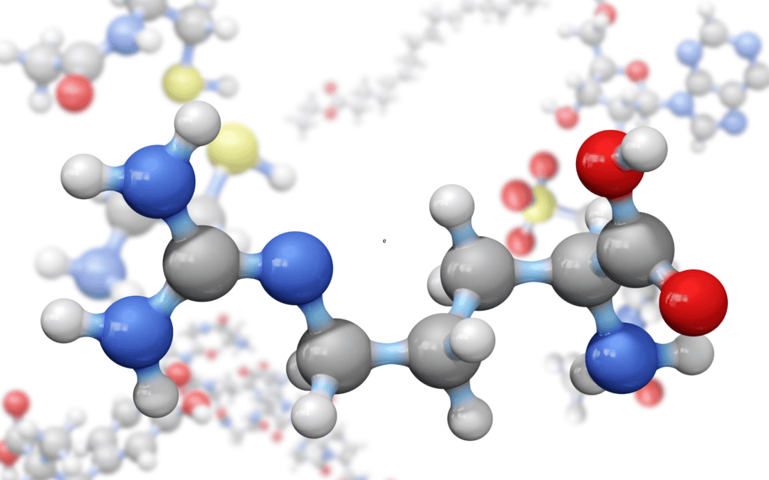
A peptide is a short chain of amino acids—essentially a fragment of a protein. Peptides serve as signaling molecules in the body, acting as hormones, neurotransmitters, immune messengers, and regulators of tissue repair.
BPC-157 (Body Protection Compound-157) is a synthetic 15-amino-acid peptide derived from a naturally occurring protein fragment found in human gastric juice. It is made entirely in a lab using solid-phase peptide synthesis—a chemical process that builds peptides one amino acid at a time on a polymer resin.
Important: BPC-157 is not derived from natural tissue. It is 100% synthetic, and the finished material depends entirely on the quality of the manufacturing process.
2. FDA Regulatory Status
The FDA’s position is unambiguous:
BPC-157 is not an approved drug for any medical condition.
It cannot be legally compounded (it appears on FDA’s “not allowed for compounding” list under 503A/503B).
It cannot be sold as a dietary supplement (not a legal dietary ingredient).
Its use in humans outside an FDA-authorized clinical trial is illegal.
Despite this, some clinics administer BPC-157 obtained from “research peptide” suppliers, offshore pharmacies, or gray-market manufacturers. These practices violate the Federal Food, Drug, and Cosmetic Act, even if enforcement has been sporadic.
3. Evidence for Use in Arthritis or Orthopedic Conditions
Bottom line: there is no high-quality evidence that BPC-157 helps arthritis in humans.
A. What exists in the scientific literature?
1. Preclinical (animal) data
Rodent studies suggest BPC-157 may:
Promote blood vessel growth
Enhance tendon fibroblast activity
Reduce inflammation
Protect gastrointestinal tissues
However, these findings:
Use doses far above any human equivalent
Use controlled laboratory injuries
Do not predict human efficacy
2. Human clinical evidence
There are only two categories of human data—and neither supports its use in arthritis.
(a) Gastrointestinal trials (Ulcerative Colitis)
A Croatian research group conducted:
A phase I safety study in healthy men
A phase II trial of a rectal BPC-157 formulation for mild–moderate ulcerative colitis
These are only available as abstracts and drug summaries, not as full peer-reviewed publications with methods, statistics, or adverse event profiles.
They do not involve musculoskeletal diseases.
(b) Single small case series in knee pain
A peer-reviewed article (Lee et al., Alternative Therapies in Health & Medicine, ~2021) describes:
12 patients with chronic knee pain
Treated with intra-articular BPC-157, often combined with TB-500
7 of 12 reported symptom improvement lasting several months
This study has major limitations:
No control group
No blinding
No standardized outcomes (no KOOS, WOMAC, or imaging)
Very small sample size
Mixed interventions
Conclusion: This study cannot demonstrate true benefit. At best, it is hypothesis-generating, and at worst, the findings may simply reflect placebo effects or biased reporting.
3. What do systematic reviews say?
Independent medical reviews and regulatory analyses uniformly state:
No controlled trials exist for arthritis, tendon injuries, or musculoskeletal pain.
No evidence-based dosing, delivery method, or safety profile is available.
Human efficacy remains unproven.
In short:There is no credible clinical evidence that BPC-157 treats arthritis or orthopedic conditions in humans.
4. Manufacturing, Purity, and Safety Concerns
Why manufacturing matters
BPC-157 is made using a complex chemical process involving:
In pharmaceutical-grade production, these chemicals are removed to trace levels through validated purification steps and quality control testing.
But “research-grade” peptide vendors selling to clinics do not follow pharmaceutical manufacturing standards.
This creates serious safety concerns:
A. Toxic solvent residues
DMF (dimethylformamide)
Known liver toxin
Associated with reproductive toxicity
Must be removed to extremely low limits in GMP drugs
DCM (dichloromethane / methylene chloride)
Probable human carcinogen
Central nervous system depressant
Dangerous even in small amounts if injected
TFA (trifluoroacetic acid)
Corrosive
Can remain in peptides as unmeasured TFA salt
Pharmaceutical products often convert TFA salts to safer counterions
Without validated HPLC, mass spectrometry, and residual solvent testing, patients may be injected with harmful chemical contaminants.
B. Unknown purity and identity
Peptides from unregulated sources may contain:
Incorrect amino acid sequence
Truncated peptides
Impurities from incomplete synthesis
Other peptides entirely
Particulate matter from resin or filters
Independent labs have repeatedly shown that many online peptides are mislabeled or impure.
C. Sterility and endotoxin concerns
Most “research peptides” are not sterile, and powder sterilization is nearly impossible without degrading the peptide. Reconstituting a non-sterile lyophilized powder with bacteriostatic saline does not sterilize it.
Risks include:
Contamination with bacteria or fungi
Endotoxin (bacterial cell wall fragments) that remain even after sterilization
Severe inflammatory reactions or sepsis
D. No validated dosing, pharmacokinetics, or long-term safety
There is no human pharmacokinetic data for injectable BPC-157:
How long it lasts in the body
What tissues it reaches
Whether it accumulates
How it is metabolized or excreted
Long-term risks cannot be assessed without such information.
5. Practical Conclusions for Arthritis and Orthopedic Use
1. Evidence of benefit in humans does not exist.
No randomized trials
No controlled studies
No validated imaging or outcome measures
Only one very small, biased case series
2. Safety is unknown—and potentially problematic.
Manufacturing is unregulated
Solvent residues may remain
Purity is unverified
Sterility is not assured
3. Clinics offering BPC-157 are doing so outside the law.
This increases the risk that:
Products may be contaminated or mislabeled
Adverse events may not be monitored or reported
Patients may be misled about efficacy
4. The scientific and regulatory communities do not support its medical use.
Anti-doping agencies, regulatory bodies, and independent medical reviews unanimously classify BPC-157 as:
Unapproved
Unproven
Potentially unsafe
6. Final Takeaway
BPC-157 is a promising laboratory molecule, not a proven medical treatment.
For arthritis and musculoskeletal pain, the data can be summarized in one sentence:
There is no credible human evidence that BPC-157 works, and real risks exist due to poor-quality manufacturing and illegal distribution.
Until properly designed, peer-reviewed human clinical trials are completed—and an FDA-regulated production pathway exists—clinicians should remain cautious and avoid its clinical use in patients.
Tendon injuries and chronic tendon degeneration — often called tendinopathy — are common yet stubbornly difficult to treat. From athletes pushing through pain to older adults coping with age-related wear, tendon issues can severely limit movement and quality of life. Traditional approaches like rest, physical therapy, anti-inflammatory medications, or surgery remain standard, but many people still struggle with lingering pain and functional limitations. Thankfully, emerging regenerative medicine approaches offer new hope — and a fundamentally different way of thinking about healing.
What Makes Tendons Hard to Heal?
Unlike other tissues, tendons have a poor blood supply, limiting their natural healing ability. When injured, tendons often form scar tissue rather than regenerating healthy tendon fibers, which can lead to chronic pain and repeated injury cycles. Aging, repetitive strain, and certain systemic conditions can worsen this process, making degeneration a long-lasting problem.
Regenerative Medicine: Healing Over Scarring
Regenerative medicine offers therapies that go beyond simply reducing symptoms. Instead, these treatments aim to stimulate the body’s own healing mechanisms, potentially leading to more complete tissue repair.
Here are some of the most studied and promising options:
1. Platelet-Rich Plasma (PRP) Therapy
PRP involves drawing a patient’s own blood, concentrating the platelets, and then injecting this platelet-rich plasma into the injured tendon. Platelets release growth factors that may help stimulate tendon cell activity, reduce inflammation, and support repair. PRP is one of the most widely used regenerative treatments for tendon injuries and is often offered as an outpatient procedure with minimal downtime.
2. Stem Cell-Based Therapies
Mesenchymal stem cells (from bone marrow or adipose tissue) are being researched for their ability to differentiate into tendon-like cells and secrete healing signals. Early studies suggest they could help modulate inflammation and support regeneration. Although clinical evidence continues to evolve and large, high-quality trials are still needed, many researchers view stem cells as a key frontier in tendon repair science.
3. Next-Generation Blood Factor Treatments
Beyond traditional PRP, newer approaches aim to isolate specific growth factors or concentrate regenerative signals more precisely. Plasma-Derived Factor treatments (like PDF-FD) extract and concentrate key proteins from blood, then deliver them directly to the injured site — potentially enhancing healing responses.
Other Frontiers: Biomaterials & Tissue Engineering
Researchers are also looking at biomaterials, scaffolds, and guided tissue engineering as ways to support tendon regeneration structurally and biologically. These approaches use engineered materials to provide a framework for tendon cells to grow — which, in theory, could rebuild tissue more like the original tendon. Although many of these are still in early stages, they represent exciting future directions.
What to Know Before Considering Regenerative Treatments
Evidence varies: While some regimens are backed by clinical research, others are still experimental. The strength of evidence for effectiveness differs across therapies, and not all treatments are universally accepted by mainstream medical societies.
Regulation and safety: Some regenerative therapies are regulated, others are offered at clinics without standardized oversight. Discuss risks, benefits, and alternatives with a qualified provider.
Not a magic bullet: Regenerative treatments are most effective when combined with physical therapy, lifestyle changes, and proper loading programs to support tendon health.
Conclusion
Tendon injuries and degeneration don’t have to be resigned to “rest and wait.” The emerging world of regenerative medicine showcases promising paths to enhance healing, stimulate regeneration, and restore function — even for chronic tendon issues. As research advances and clinical evidence grows, these biologic solutions may reshape how we approach tendon care in the years ahead.
Joint degeneration doesn’t happen overnight. It’s a slow, progressive process — but the earliest warning signs are often subtle enough that many people shrug them off as “getting older” or “overdoing it.” Identifying these changes early can make all the difference in treatment outcomes, mobility, and long-term joint health. And thanks to advances in regenerative medicine, patients now have powerful options that can protect, repair, and even restore damaged tissue before the problem becomes severe.
Below, we break down the early symptoms to watch for and explore how modern regenerative therapies can help interrupt the degenerative cascade.
What Early Joint Degeneration Looks Like
1. Persistent Morning Stiffness
If you feel “rusty” or tight first thing in the morning — especially in the knees, hips, or spine — this can indicate early cartilage wear or inflammation. Stiffness that improves as you move around is a classic hallmark of early degenerative changes.
2. Clicking, Popping, or Grinding
Noisy joints aren’t always dangerous, but when those sounds are paired with discomfort, swelling, or instability, they may signal roughened surfaces or early cartilage breakdown.
3. Pain With Activity That Improves With Rest
You might feel fine at rest but experience dull aching during:
Walking up or down stairs
Long periods of standing
Squatting or lifting
High-impact workouts
This “use-related pain” often shows up years before advanced arthritis.
4. Swelling or Warmth Around the Joint
Mild inflammation is one of the earliest internal reactions to joint stress or degeneration. Even occasional swelling after activity can indicate chronic irritation inside the joint.
5. Reduced Range of Motion
Maybe your knee doesn’t bend the way it used to, or your shoulder feels tight when reaching overhead. Loss of flexibility is often one of the first measurable signs of joint deterioration.
6. Fatigue or Weakness in the Joint
Muscles surrounding a degenerating joint often work overtime to compensate, which can create fatigue, shakiness, or weakness during routine tasks.
Why Early Action Matters
Once cartilage has worn away significantly, it cannot regenerate on its own — and late-stage arthritis often requires aggressive treatments like joint replacement. Early intervention, however, can slow or even stop the degenerative cycle. The goal is to reduce inflammation, restore stability, and stimulate natural repair pathways before more permanent damage occurs.
This is where regenerative medicine shines.
How Regenerative Therapies Can Help
Platelet-Rich Plasma (PRP)
PRP concentrates your body’s own growth factors to:
Reduce inflammation
Promote tissue repair
Slow cartilage breakdown
Improve joint lubrication
Great for early osteoarthritis, tendon irritation, and chronic joint strain.
Orthobiologics
These treatments use biologically active cells and proteins to enhance healing in damaged tissue. Orthobiologics can improve structural stability, reduce inflammation, and support long-term joint health.
Cell–Based Therapies
These therapies may help stimulate new tissue formation in joints that show early degenerative changes. They can support:
Cartilage protection
Improved mobility
Reduced pain
Enhanced shock absorption
Especially useful for knees, hips, shoulders, and spine-related degeneration.
Hyaluronic Acid (Viscosupplementation)
A lubrication-boosting gel injected into the joint to:
Improve glide
Reduce pain
Support smoother movement
Works well for mild to moderate osteoarthritis.
Lifestyle + Regenerative = Long-Term Prevention
The best outcomes happen when regenerative therapies are paired with:
Strength training
Weight management
Physical therapy
Anti-inflammatory nutrition
Activity modification
Together, these strategies help reinforce joint support structures and reduce ongoing stress.
When to Seek Evaluation
You don’t need severe pain to justify seeing a specialist. In fact, the sooner you understand what’s happening inside your joints, the more options you have to preserve their health.
Seek evaluation if you notice:
Recurring stiffness
Activity-related pain
Early swelling
Clicking or grinding
Weakness or instability
A simple exam — sometimes paired with X-ray or ultrasound — can determine your level of joint degeneration and whether regenerative therapies are appropriate.
Final Thoughts
Joint degeneration doesn’t have to be inevitable or irreversible — especially when it’s caught early. Modern regenerative options offer patients a chance to protect and restore joint health naturally, often delaying or avoiding more invasive procedures down the line.
If you’re starting to notice the early signs, now is the perfect time to explore your options and take proactive steps to keep your joints strong, mobile, and pain-free for years to come.